Amino acids and proteins
Unit 2 · Chemical and Cellular Basis of Life
Amino acid structure, peptide bonds, the four levels of protein structure, denaturation, and the functions of proteins; the biuret test.
Amino acids
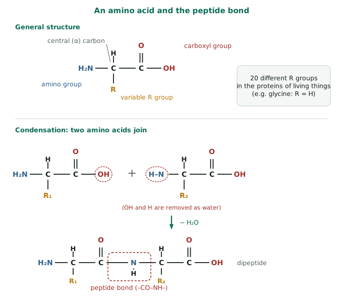
Proteins are polymers of amino acids. All amino acids have the same basic structure: a central ($\alpha$) carbon atom bonded to
- an amino group ($\text{-NH}_2$),
- a carboxyl group ($\text{-COOH}$),
- a hydrogen atom, and
- a variable R group (side chain).
Twenty different R groups occur in the proteins of living things. The R group may be small (glycine, R = H), polar, charged (acidic or basic) or non-polar, and it determines the properties of the amino acid. In solution, at neutral pH, an amino acid exists as a zwitterion ($\text{-NH}_3^+$ and $\text{-COO}^-$), so it can act as a buffer, accepting or giving $\text{H}^+$. Humans cannot make about 9 of them (essential amino acids) and must obtain them in the diet.
The peptide bond
In a condensation reaction, the carboxyl group of one amino acid joins the amino group of the next, releasing water and forming a peptide bond ($\text{-CO-NH-}$). Two amino acids form a dipeptide, and many form a polypeptide. A protein consists of one or more polypeptide chains. Hydrolysis (digestion by proteases) breaks the peptide bonds.
Levels of protein structure
- Primary structure: the sequence of amino acids in the polypeptide, coded by the gene. A single change in sequence can change the whole protein (for example, sickle-cell haemoglobin has valine instead of glutamic acid at one position).
- Secondary structure: the polypeptide folds locally into an α-helix or a β-pleated sheet, held by hydrogen bonds between the $\text{C=O}$ and $\text{N-H}$ groups of the backbone.
- Tertiary structure: the whole chain folds into a specific three-dimensional shape through interactions between R groups: hydrogen bonds, ionic bonds, disulfide bridges (covalent, between two cysteines) and hydrophobic interactions (non-polar R groups cluster in the core).
- Quaternary structure: two or more polypeptides assemble into one functional protein (haemoglobin has four chains; insulin has two). Some also have a non-protein prosthetic group, such as the haem group in haemoglobin.
Types and functions
| Fibrous | Globular | |
|---|---|---|
| Shape | Long, rope-like chains | Compact, rounded |
| Solubility | Insoluble | Soluble |
| Role | Structure and strength | Metabolic: enzymes, transport, hormones |
| Examples | Collagen (tendons, skin), keratin (hair, nails), fibroin (silk) | Enzymes, haemoglobin, insulin, antibodies |
Other functions: transport across membranes (carrier and channel proteins), movement (actin and myosin), defence (antibodies), hormones (insulin), storage (casein in milk, albumin in egg) and receptors.
Denaturation
The shape of a protein is maintained by weak bonds. High temperature, extreme pH, heavy metals and organic solvents break these bonds, so the protein loses its three-dimensional shape (denaturation). Because function depends on shape, an enzyme or antibody that is denatured stops working. Boiling an egg is a familiar example of irreversible denaturation.
Biuret test
Add sodium hydroxide, then a few drops of dilute copper(II) sulfate to the sample. A purple (lilac) colour indicates peptide bonds, so protein is present; it stays blue if there is none.
Deshapriya Senevirathne. B.Sc. (Hons) Biology Special, University of Peradeniya — graduated with a class pass. All the content and lessons in A/L Biology on Paadama are reviewed by Deshapriya Senevirathne, and the lessons are taught by him.